 Take Home Points
- Stress cardiomyopathy looks like ACS/STEMI, with patient presenting with chest pain, dyspnea or maybe syncope. It looks like ACS and should be treated as such until you prove to yourself it’s not.
- Classic patient is an older woman with chest pain or syncope after a stressful event.
- Bedside echo will show left ventricular dysfunction with one of a variety of patterns of wall motion abnormality. The most common is apical, but there are also variant patterns including mid-ventricular, basal, focal and global.
- Watch for QTc prolongation as this could precipitate an arrhythmia. Be sure to stop all QT prolonging meds and replete magnesium
- Consider the differential in the patient who has cardiogenic shock because the treatment differs. Avoid catecholamines and if you need inotropic support use dobutamine or dopamine. Look for evidence of left ventricular outflow tract (LVOT) obstruction, as this should be treated like a hypertrophic cardiomyopathy with beta blockers rather than inotropes.
REBEL Core Cast 20.0 – Takotsubo’s Cardiomyopathy
Background
- Stress cardiomyopathy goes by many names including takotsubo cardiomyopathy, apical ballooning syndrome, broken heart syndrome, and stress-induced cardiomyopathy.
- The patient has transient regional systolic dysfunction of the left ventricle.
- Clinically, this mimics a myocardial infarction, but when an angiogram is performed there is no evidence of obstructive coronary artery disease or acute plaque rupture.
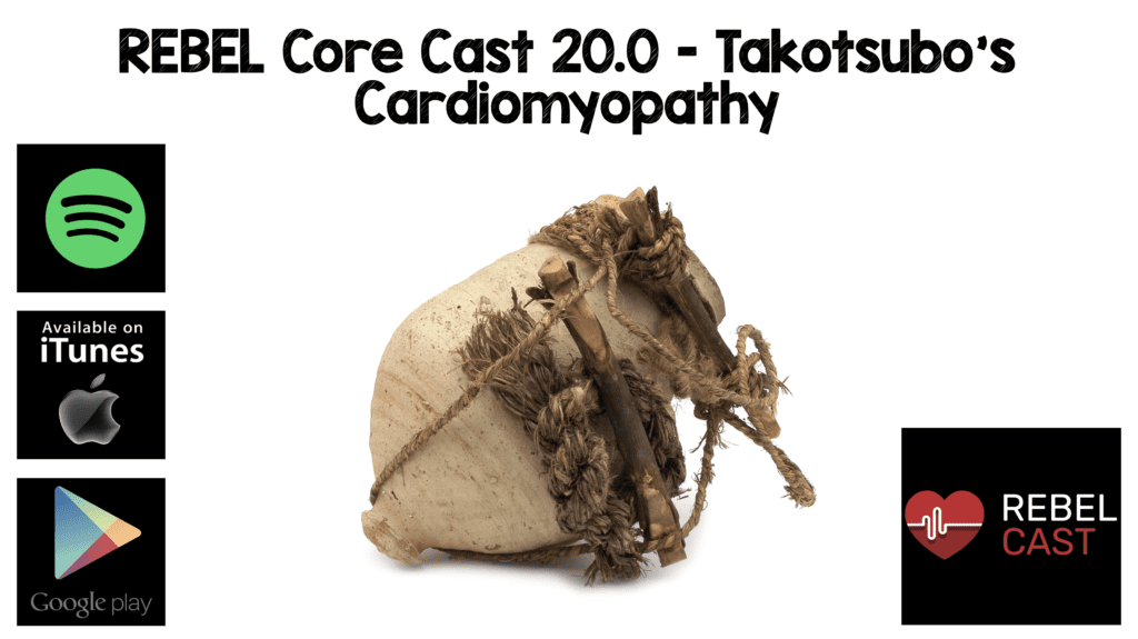
- The term “takotsubo” is from the Japanese name for an octopus trap, which has a shape that is similar to the systolic apical ballooning appearance of the left ventricle which is seen in the most common form of this disorder.
Presentation
- These patients will present looking very similar to a classic acute coronary syndrome, with chest pain and shortness of breath.
- Additionally, according to the International Takotsubo Registry study approximately 7.7% will present with syncope.
- Most common in elderly female patients.
EKG Findings
- EKG is highly variable.
- Patients may present with normal ECG up to 11% of the time.
- Could have ST/T wave changes, ST elevation, transient LBBB or arrhythmias.
- The most characteristic takotsubo ECG feature is a prolongation of QT/QTc
Lab Findings
- May have elevated troponin and BNP.
Echo Findings (https://www.ultrasoundoftheweek.com/uotw-74/)
- Usually will have decreased ejection fraction ~40%
-
Wall motion abnormalities in one of a few specific patterns:
- Apical – Systolic apical ballooning of the LV
- Mid-ventricular – Ventricular hypokinesis of the mid-ventricle with relative sparing of the apex
- Basal – Hypokinesis of the base and sparing of the mid-ventricle and apex (called reverse or inverted Takotsubo)
- Focal – Rare variant with dysfunction of an isolated segment
Treatment
- Always follow ACS/STEMI treatment protocol until this diagnosis is proven.
-
Monitor the QTc interval and arrhythmia.
- Stop QT prolonging medications
- Replete electrolytes
-
Cardiogenic shock
- If the patient has pump failure and no LVOT obstruction, treat with inotropes like dobutamine and dopamine
-
If the patient has pump failure and evidence of LVOT obstruction, treat similar to hypertrophic cardiomyopathy
- Inotropes may worsen the obstruction.
- Treat with beta blockers.
- If vasopressors are needed, consider phenylephrine
Resources
- WikEM – Takotsubo Cardiomyopathy
- BBC.com – The Real Life Diseases That Spread the Vampire Myth
- Dawson, DK. Acute stress-induced (takotsubo) cardiomyopathy. Heart. 2018 Jan;104(2):96-102. doi: 10.1136/heartjnl-2017-311579. Epub 2017 Aug 20. PMID: 28824005
- Meigh, K. et al. Takotsubo Cardiomyopathy in the Emergency Department: A FOCUS Heart Breaker. Clin Pract Cases Emerg Med. 2018 Apr 5;2(2):158-162. doi: 10.5811/cpcem.2018.2.37291. eCollection 2018 May. PMID 29849274
- Rigor, J et al. Porphyrias: A clinically based approach. Eur J Intern Med. 2019 Sep;67:24-29. doi: 10.1016/j.ejim.2019.06.014. Epub 2019 Jun 27. PMID: 31257150
Post Peer Reviewed By: Salim R. Rezaie, MD (Twitter: @srrezaie)
The post REBEL Core Cast 20.0 – Takotsubo’s Cardiomyopathy appeared first on REBEL EM - Emergency Medicine Blog.
