 Take Home Points
- SBO should be considered in all patients presenting with abdominal pain particularly if they have a prior abdominal surgical history
- Patients with SBO often have non-specific signs and symptoms. There is no history or physical exam feature that rules out the disease
- Lactate elevation is a late finding in SBO. A normal lactate does not rule out the diagnosis
- Plain X-rays perform poorly in making or ruling out the diagnosis. CT is the most widely accepted imaging modality but US (both formal and ED) has better performance characteristics
- Patients with SBO should have an emergent surgical consultation and treatment should start with good supportive care (IV fluids, electrolyte repletion, antiemetics)
- Patients with hypotension, hypoperfusion or frank sepsis from an SBO should be aggressively resuscitated and optimized for operative management
REBEL Core Cast 94.0 – SBO
Definition: Obstruction of the intestines such that bowel contents are unable to pass through the small bowel into the large bowel
- Mechanical Obstruction: presence of a physical barrier to the passage of bowel contents.
-
- Examples: adhesions, neoplasms, inflammatory disease (i.e. Crohn’s), hernias, intussusception, parasitic infections and foreign bodies
- Simple Obstruction: Obstruction occurs at a single point in the bowel
- Closed-loop obstruction: Obstruction at two locations creating a segment of bowel with proximal and distal compromise of blood flow
-
- Functional (neurogenic) Obstruction: Obstruction resulting from disruption of normal peristalsis in the GI tract in the absence of a mechanical obstruction (adynamic ileus). Examples include post-operative, hypokalemia and opiate use
Pathophysiology
- Dilation of the bowel proximal to the obstruction
- Results in accumulation of intestinal secretions and partially digested materials
- Stimulates peristalsis resulting in early loose bowel movements as well as nausea and vomiting
- Bowel wall becomes edematous
- Decreased absorption capacity leading to further dilation
- Decreased venous return and, ultimately, decreased arterial flow
- Bacterial overgrowth occurs as a result of stasis
- Translocation of bacteria (notably E.Coli) into the bloodstream can occur due to increased bowel wall permeability
- Extensive fluid losses can result from transudative fluid loss into the bowel lumen
- Recurrent vomiting can lead to metabolic alkalosis, electrolyte abnormalities and hypovolemia
-
Closed Loop Pathophysiology
- Intraluminal pressure increases more rapidly due to inability of bowel contents to move in retrograde fashion
- Venous and arterial compromise can occur more rapidly leading to intestinal ischemia and infarction
- Infarction and necrosis of the bowel can lead to perforation with resulting peritonitis and sepsis

Presentation (Taylor 2013)
- History
- Abdominal pain
- Often crampy in nature
- Crescendo-decrescendo pattern
- Abdominal distension
- Nausea/Vomiting (more pronounced with proximal obstructions)
- Constipation
- Obstipation (inability to pass gas- typically a late finding)
- Prior abdominal surgery or radiation treatments common
- Abdominal pain
- Physical Examination
- Early findings
- Generalized tenderness to palpation
- Decreased bowel sounds or high pitched “tinkling”
- (+) LR Abnormal Bowel Sounds: 6.33
- (-) LR Abnormal Bowel Sounds: 0.27
- Late findings
- Fever
- Abdominal distension
- (+) LR: 5.64 – 16.8
- (-) LR: 0.34 – 0.43
- Absent bowel sounds
- Peritoneal signs (i.e. rebound and guarding)
- Early findings
Diagnostics
- Laboratory Tests
- Commonly ordered lab tests (i.e. WBC) are non-sensitive and non-specific
- Serum lactate
- Elevated in cases of bowel ischemia or infarction
- Elevation typically a late finding
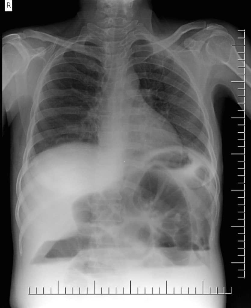
- Abdominal X-ray
- Findings
- Dilated loops of small bowel proximal to the obstruction
- Visible valvulae conniventes (mucosal folds of the small intestine)
- Air-fluid levels
- String-of-beads sign (small pockets of gas within a fluid-filled small bowel)
- Advantages: Rapidly performed, non-invasive, can be performed bedside in unstable patients
- Disadvantages: Poor sensitivity and specificity
- Test Characteristics (Taylor 2013)
- (+) LR: 1.6 (1.1 – 2.5)
- (-) LR: 0.43 (0.24 – 0.79)
- Findings
-

CT Scan with Fluid Filled, Dilated Loops of Bowel CT Scan
- Findings
- Dilated loops of small bowel > 2.5 – 3 cm (measure outer wall to outer wall)
- Collapsed loops of bowel distal to the obstruction
- Intestinal Ischemia/Infarction
- Thickened bowel wall
- Increased attenuation of the bowel wall
- Pneumatosis intestinalis (non-specific)
- Portal venous gas (non-specific)
- Advantages
- Increased sensitivity and specificity to plain radiographs
- Often identifies cause of obstruction
- Can identify alternate pathology or cause of symptoms
- Disadvantages
- Takes more time
- Requires patient to leave department (may not be feasible if patient unstable)
- Test Characteristics (5 – 10 mm slice) (Taylor 2013)
- (+) LR: 3.6 (2.3 – 5.4)
- (-) LR: 0.18 (0.09 – 0.35)
- Findings
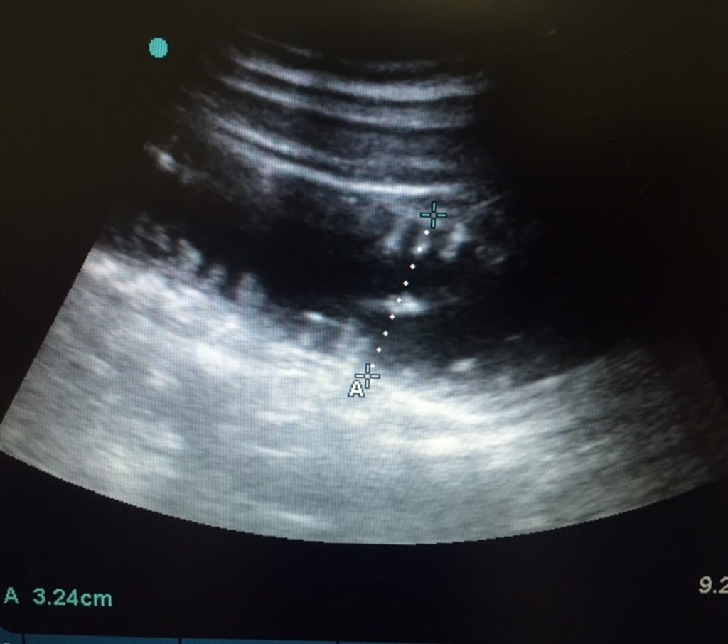
- Ultrasound
- Findings
- Dilated loops of bowel (diameter > 2.5 cm) – most sensitive and specific
- Decreased peristalsis and retrograde peristalsis (to and fro sign or “whirling”)
- Bowel Infarction/Ischemia
- Fluid filled bowel with extra-luminal free air
- Bowel wall thickening (> 3 mm)
- Absence of peristalsis
-

US With Dilated Loops of Bowel Advantages
- Non-invasive
- Can be rapidly performed at the patient’s bedside
- Excellent test characteristics
- Disadvantages
- Dependent on operator skill
- Patient habitus may limit quality of images
- Does not clearly delineate transition point
- May not be accepted by consulting services for definitive diagnosis
- Test Characteristics (Taylor 2013)
- Formal US
- (+) LR: 14.1 (3.6 – 55.6)
- (-) LR: 0.13 (0.08 – 0.20)
- ED US
- (+) LR: 9.5 (2.1 – 42.2)
- (-) LR: 0.04 (0.01 – 0.13)
- Formal US
- Findings
Immediate Management:
Basics:
- Airway
- It is uncommon for patients with SBO to have airway compromise
- Patients with closed-loop obstruction can become septic and obtunded while having continued vomiting
- Circulation
- Advanced cases can develop septic shock
- Most cases will have intravascular volume depletion secondary to transudation of fluids into the bowel and decreased PO intake
- Volume expansion with isotonic fluids (Lactated ringers, 0.9% saline or balanced solutions) indicated
- Electrolyte repletion as necessary (fluid loss will be accompanied by electrolyte loss)
- Antiemetics: may be helpful in those with partial SBO and in those not actively vomiting
Directed Management
- Surgical Consultation
- Surgical exploration indicated (Maung 2012)
- Patients with evidence of clinical deterioration
- Patients with generalized peritonitis
- CT findings with bowel ischemia or infarction
- Non-operative patients will require serial abdominal examinations and consideration for operative management if the clinical picture changes
- Surgical exploration indicated (Maung 2012)
- Nasogastric Tube (NGT) Placement
- Traditional teaching argues for the use of NGT in all patients with SBO but literature to defend this approach is minimal (Paradis 2014)
- Resolution of nausea/vomiting with antiemetics may obviate the need for an NGT
- Indications for NGT placement
- Intractable vomiting
- Severe abdominal distension
- Altered mental status or other aspiration risk
- Antibiotics
- Theory is that bacterial translocation from the gut will be increased due to bowel wall edema
- Limited data to support administration in all cases (Maung 2012)
- Broad-spectrum antibiotics should be administered in patients with signs of hypoperfusion and sepsis
- Disposition
- All patients should be admitted to a surgical service
- Limited evidence demonstrates lower mortality and better outcomes in SBO admitted to surgical service (Oyasiji 2010)
- Patients more likely to get operative intervention earlier upon decompensation
- Patients with closed loop obstructions or signs of ischemia/infarction should be admitted to an ICU setting
- All patients should be admitted to a surgical service
Take Home Points
- SBO should be considered in all patients presenting with abdominal pain particularly if they have a prior abdominal surgical history
- Patients with SBO often have non-specific signs and symptoms. There is no history or physical exam feature that rules out the disease
- Lactate elevation is a late finding in SBO. A normal lactate does not rule out the diagnosis
- Plain X-rays perform poorly in making or ruling out the diagnosis. CT is the most widely accepted imaging modality but US (both formal and ED) has better performance characteristics
- Patients with SBO should have an emergent surgical consultation and treatment should start with good supportive care (IV fluids, electrolyte repletion, antiemetics)
- Patients with hypotension, hypoperfusion or frank sepsis from an SBO should be aggressively resuscitated and optimized for operative management
Read More
- The Short Coat: Small Bowel Obstruction – A Likely Story?
- Radiopaedia: Small Bowel Obstruction
- 5 Min Sono: SBO
- Ultrasound Podcast: Episode 36 – Small Bowel Obstruction
- LITFL: Small Bowel Obstruction
- FOAMCast: Episode 23 – SBO and Mesenteric Ischemia
- Roline CE, Reardon RF: Disorders of the small intestine, in Marx JA, Hockberger RS, Walls RM, et al (eds): Rosen’s Emergency Medicine: Concepts and Clinical Practice, ed 8. St. Louis, Mosby, Inc., 2010, (Ch) 92: p 1216-1227.
References
- Taylor MR, Lalani N. Adult small bowel obstruction. Acad Emerg Med 2013; 20(6): 528-44. PMID: 23758299
- Maung AA et al. Evaluation and management of small-bowel obstruction: an eastern association for the surgery of trauma practice management guideline. J Trauma Acute Care Surg 2012; 73(5): S362-9. PMID: 23114494
- Paradis M. Towards evidence-based Emergency Medicine: Best BETs From the Manchester Royal Infirmary. BET 1: Is Routine Nasogastric Decompression Indicated in Small Bowel Occlusion? Emery Med J 2014; 31(3): 248-9. PMID: 24532357
- Oyasiji T et al. Small bowel obstruction: Outcome and cost implications of admitting service. Am Surg 2010; 76:687-691. PMID: 20698371
- Images
- Air Fluid Levels: Case courtesy of Dr Aditya Shetty, <a href=”https://radiopaedia.org/”>Radiopaedia.org</a>. From the case <a href=”https://radiopaedia.org/cases/28737″>rID: 28737</a>
- CT SBO Case courtesy of Dr Jeremy Jones, <a href=”https://radiopaedia.org/”>Radiopaedia.org</a>. From the case <a href=”https://radiopaedia.org/cases/6135″>rID: 6135</a>
- US SBO Case courtesy of Dr Maryam Saif AlAli, <a href=”https://radiopaedia.org/”>Radiopaedia.org</a>. From the case <a href=”https://radiopaedia.org/cases/43438″>rID: 43438</a>
|
Sensitivity |
Specificity |
(+) LR |
(-) LR |
|
|
Decr Bowel Sounds (or high pitched “tinkling) |
23 – 76% |
88 – 93% |
3.29 – 6.33 |
0.27 – 0.83 |
|
Abdominal Distension |
62 – 67% |
89 – 96% |
5.64 – 16.8 |
0.34 – 0.43 |
|
Abdominal X-ray |
75% |
66% |
1.6 |
0.43 |
|
CT Scan |
87% |
81% |
3.6 |
0.18 |
|
Radiology US |
90% |
96% |
14.1 |
0.13 |
|
ED US |
97%% |
90%% |
9.5 |
0.04 |
Post Peer Reviewed By: Salim R. Rezaie, MD (Twitter: @srrezaie)
The post REBEL Core Cast 94.0 – SBO appeared first on REBEL EM - Emergency Medicine Blog.
