 A 64 year old man with an extensive history of abdominal surgeries presents to the emergency department with abdominal pain and vomiting. Because you suspect a bowel obstruction, you bring an ultrasound machine to the bedside prior to the completion of any laboratory testing or other imaging. A curvilinear probe in the abdominal mode setting was used to scan in all four quadrants of the abdomen looking in both the sagittal and transverse planes.
Ultrasound imaging
With a curvilinear probe on the patient’s abdomen (Figure 1), the following ultrasound (US) image (Figure 2) and video (Figure 3) were obtained. The images showed dilated, fluid-filled bowel loops with thickened bowel walls, as well as minimal peristalsis. A CT scan confirmed a diagnosis of a distal small bowel obstruction (SBO).

Figure 1. Position of the curvilinear prone on the patient’s abdomen

Figure 2. Curvilinear probe used to demonstrate multiple small bowel loops and abdominal free fluid
SBO: Background
Annually, in the United States, approximately 10% of all visits to the emergency department, or 13 million patients, present with the chief complaint of abdominal pain1. Of those patients, approximately 2% are diagnosed with an SBO.2 While not a very common cause of abdominal pain, it is associated with high rates of severe complications,3 including strangulation and bowel necrosis.4,5 Bickell et al found that a delay in the diagnosis and management of an SBO was associated with a higher risk of bowel resection. Only 4% of patients appropriately managed less than 24 hours after symptom onset required resection compared with 10-14% of patients managed more than 24 hours after symptom onset.6 Emergency medicine (EM) physicians have a unique role to lower the likelihood of poor outcome in individuals with SBO as the vast majority of patients subsequently diagnosed with an SBO initially present to the ED.7
SBO: History and Physical
When a patient presents with abdominal pain, the history and physical exam can help differentiate SBO from other causes of pain. Components of the history and physical exam more commonly associated with an SBO include a history of previous surgeries, constipation, abnormal bowel sounds, and abdominal distention.3 In one study, a history of previous abdominal surgery with adhesions was seen in 75% of patients with SBO.8 Constipation and abdominal distention can also point towards the diagnosis of an SBO, but have been found to have poor sensitivities.9,10
SBO: Imaging
While the physical exam plays an important part in the initial evaluation of a suspected SBO, imaging plays a critical role in its definitive diagnosis. Multiple imaging modalities have been described to diagnose SBO, including CT, MRI, X-ray, and US.3
- CT has been found to be highly accurate for the diagnosis of SBO. Two studies using a 64 slice multidetector CT demonstrated sensitivities of 93%-96% and specificities of 93%-100% for the diagnosis of SBO.11,12 With its high accuracy, the CT scan is considered to be the gold standard for diagnosis of SBO.13
- MRI can be a useful alternative to a CT scan and has been reported to have similar accuracies when compared to a 64-slice CT scanner.14,15
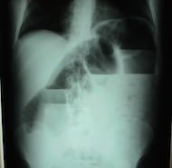
- Abdominal X-Ray (AXR) have been found to have disappointing accuracy. The only publication that used CT as the sole gold standard for SBO found AXR to have a sensitivity of 46.2% and a specificity of 66.7%.16
Although highly accurate, both the CT and the MRI have the distinct disadvantages of not being able to performed at the bedside, as well as being time consuming, more costly, and in the case of CT, carrying the side effects of radiation and possible contrast reactions. Ultrasound is a bedside testing modality that has recently arisen as a viable alternative.
SBO: What about ultrasonography?
There has been a recent explosion in research and expansion of bedside US as an imaging tool in the ED. Subsequently, US has emerged as a possible adjunct in the accurate and timely diagnosis of SBO. Limited research to date has been performed using US as a diagnostic modality for an SBO. A recent meta analysis and systemic review identified six prospective US studies, only two of which were done in the ED.3
- Unluer et al performed a prospective study that enrolled 174 patients in the ED, 90 of which eventually were given the diagnosis of an SBO.13 This study used four relatively inexperienced EM residents as operators and found their ultrasounds to have a sensitivity of 97.7% and a specificity of 92.7%.
- Jang et al enrolled 76 patients, 33 of whom were eventually diagnosed with an SBO by CT scan. They found that dilated bowel on US had a sensitivity of 91% and a specificity of 84%, while decreased peristalsis had a specificity of 98% and sensitivity of 27%.16
Ultrasound criteria for diagnosing SBO
Specific criteria used in the sonographic diagnosis of an SBO vary slightly in the medical literature, but the publications reviewed considered a fluid-filled small bowel lumen >2.5 cm to be consistent with the diagnosis of SBO.13,16–18 Fluid seen outside of the dilated loops of bowel are thought to confer a worse prognosis.19
Advantages of using ultrasound for SBO
Ultrasound is a promising adjunct to the evaluation of a patient with a suspected SBO. It can be performed rapidly and with high accuracy, even in the hands of providers with minimal training. In the study by Jang et al, EM residents with 10 minutes of didactic time and previous experience with only 5 SBO ultrasounds performed with high accuracy.16 Ultrasound is a non-carcinogenic, bedside imaging modality that has the potential to decrease costs, and may be preferred in patients with relative contraindications to CT scans. Even in patients without contraindications to CT scans, US may be used to safely and quickly identify and risk-stratify those who require further imaging versus those who can be safely discharged home. In addition, patients with recurrent episodes of SBO could potentially be managed with US as the sole imaging modality to avoid multiple and repeated dosages of ionizing radiation in the form of a CT scan.
Further research on a larger scale is needed to continue to explore the utility of bedside US as a rapid, accurate and potentially life-saving option for imaging in patients with potential small bowel obstructions, and to specifically address if a patient with US as the sole imaging modality can be managed based on lack or presence of findings suspicious for SBO.
Author information
The post Small bowel obstruction: Diagnosis by ultrasonography appeared first on ALiEM.

