
A 32 year-old male presents to the Emergency Department after he felt a “pop” in his posterior-medial calf while playing tennis. He was able to ambulate but had pain with plantar flexion and was unable to continue playing tennis. What is your differential diagnosis? What physical exam maneuver would you perform? What findings would you expect on physical exam? What is the diagnosis based on ultrasound images? What is your management in the emergency department?
")

Figure 1: Case courtesy of Dr Chris O’Donnell, Radiopaedia.org
The differential diagnosis for a patient who experiences acute pain in the posterior lower leg with activity includes Achilles tendon rupture, gastrocnemius strain, soleus strain, ruptured Baker’s cyst, deep vein thrombosis, and plantaris rupture.

Figure 2: Muscles of the lower leg. OpenStax College
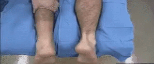
The patient’s history is concerning for potential achilles tendon rupture. To identify this injury, the astute practitioner would perform the Thompson Test (abnormal left, normal right). The Thompson Test has a sensitivity of 96% and specificity of 93% in a complete Achilles tendon rupture [1]. A negative Thompson test does not completely rule out an Achilles injury, especially clinically significant partial injuries. If clinical suspicion remains high, the patient should be treated as if they have a tendon rupture with a walking boot and heel lift, so the foot is in plantar flexion. Ultrasound of the tendon can also be useful to assist with the diagnosis.
Upon physical examination, you might find tenderness to palpation of the plantaris muscle and full strength in dorsiflexion and plantarflexion [2]. If the patient does not have achilles tendon injury they are likely to have a negative Thompson test. The plantaris plays a proprioceptive role more than a strength role, so there may not be any noticeable discrepencies in motor function.
The ultrasound images show a plantaris tendon rupture as indicated by the hypoechoic region between the gastrocnemius and soleus, in addition to the lack of normal fibrous structure of normal tendon. The plantaris is a vestigial muscle, absent in up to 20% of limbs, that lies between the gastrocnemius and soleus and contributes to plantar flexion [3]. Plantaris rupture or sometimes referred to as “tennis leg” however this term is used interchangeably for numerous pathologies; we recommend referring to the anatomic anatomy to describe the injury.

Figure 3: annotated ultrasound of plantaris rupture. Green – Medial gastrocnemius Red – Plantaris Blue – Soleus
Given the plantaris is a vestigial muscle, its rupture does not result in loss of function. Patients are treated symptomatically and can gradually return to activity as tolerated. The goal of ED treatment is pain-free ambulation which can be achieved with a walking boot and crutches if needed. Follow up with a Sports Medicine physician in 1-2 weeks.
References
- Maffulli N. The clinical diagnosis of subcutaneous tear of the Achilles tendon. A prospective study in 174 patients. Am J Sports Med, 1998. PMID: 9548122
- Spina AA. The plantaris muscle: anatomy, injury, imaging, and treatment. J Can Chiropr Assoc. 2017;51(3):158-163. PMID: 17885678
- Rohilla S, Jain N, Yadav R. Plantaris rupture: why is it important? BMJ Case Rep, 2013. PMID: 23345486
Author information

The post A Pop in the Calf – Plantaris Rupture appeared first on ALiEM.